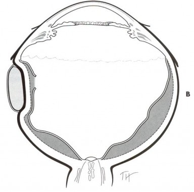

Scleral Buckling SurgeryĬonsidered the traditional surgery for retinal detachments, scleral buckling is performed using local, and in some cases, general anesthesia. There are three types of surgery for retinal detachment: 1) scleral buckling surgery, 2) pneumatic retinopexy, and 3) vitrectomy. Without surgery, vision will almost always be completely lost. If a detachment becomes too large for the laser treatment or cryotherapy alone, surgery is normally necessary to reattach the retina. If a retinal detachment or tear is small, it may be repaired using laser surgery, cryotherapy (freezing an area), or a combination of the two.

The retinal surgeon will decide the most effective treatment for a retinal detachment, hole or tear. What is the treatment for a retinal detachment, a retinal hole or a retinal tear? Left untreated, in most cases, after a retinal detachment starts, the entire retina will eventually detach and all useful vision in that eye will be lost.

Frequently, detachments begin with loss of peripheral vision and patients may notice a dark shadow, or a veil, coming from one side, above or below. This collection of fluid causes a lifting action and markedly disturbs the vision. This typically happens when liquefied vitreous fluid passes through a small tear in the retina and collects behind the retina. Retinal detachments, holes and tears are serious conditions that require immediate attention.Ī retinal detachment occurs when the retina separates from the back wall of the eye. Laser surgery is frequently performed in the office of the surgeon, but it is also utilized during other complicated retinal procedures in an outpatient surgical area. The body responds to heal the area and in so doing, a seal is created. As the light reaches the problem area to be treated, it begins to heat up. The laser generates a light source that is directed through a special contact lens or ophthalmoscope. 950 Town Center Dr.Retinal laser surgery is used to treat a variety of problems including retinal tears, retinal holes, small retinal detachments and leaking blood vessels in the back of the eye.
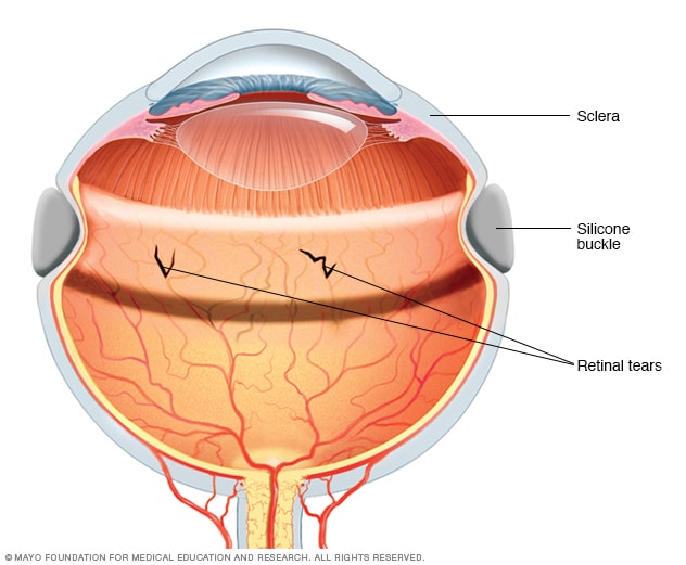
 1205 High Street | Millville, NJ | 08332. 1802 Haddonfield Berlin Road | Cherry Hill, NJ | 08003. 423 Clements Bridge Road | Barrington, NJ | 08007. 225 Sunset Road | Willingboro, NJ | 08046. If you would like to make an appointment, call us 609.877.2800 or EMail us. If a gas bubble was placed, there are specific post-surgical instructions that must be followed. Recovery time varies from person to person and can be anywhere from two to eight weeks. The gas bubble acts as a shield to prevent any further fluid from accumulating under the retina during the healing process. At this point a gas bubble may be placed in the eye. Subretinal fluid is drained and the tears or holes in the retina are sealed. The buckle usually remains in place permanently. It is secured around the white of the eye-the sclera. Reducing the eye’s diameter creates slack and eliminates the tugging and pulling from the fluid that was detaching the retina.Ī scleral buckle is a piece of silicone, semi-hard plastic that surrounds and secures the eye like a belt. The buckle is tightened around the eye to reduce the diameter of the eye. How does a Scleral Buckle Work?Ī small buckle is placed around the eye under the eye muscles. This type of retinal detachment can be caused by injury or trauma to the eye, tumors in the eye, or diseases that cause inflammation inside the eye. The most common causes of exudative retinal detachment are leaking blood vessels or swelling in the back of the eye. Other causes are eye diseases, eye infections, and swelling of the eye.Įxudative retinal detachment is also caused by fluid building up behind the retina, but there are no retinal tears or holes in this type of retinal detachment. The most common cause of tractional retinal detachment is diabetic retinopathy in which blood vessels become damaged and cause scarring. Tractional retinal detachment occurs when scar tissue grows on the retina pulling it away from the back of the eye. The tear or hole allows the gel-like fluid in the center of the eye to seep behind the retina where it pushes the retina away from the back of the eye causing it to detach. This type of detachment happens in 90% of all cases of retinal detachment. Rhegmatogenous retinal detachment is the most common type of retinal detachment and it happens when there is a small tear or hole in the retina. Other surgeries to reattach the retina are pneumatic retinopexy, vitrectomy, or a combination of scleral buckling and vitrectomy. The first scleral buckle surgery to fix a retinal detachment was performed in 1953 and with a few refinements and modifications over the years it remains a reliable and effective way to reattach a detached retina.


 0 kommentar(er)
